EPILEPSY & SEIZURES SERIES • PART ONE BY STEVEN M. WOLF, MD & PATRICIA MCGOLDRICK, RN
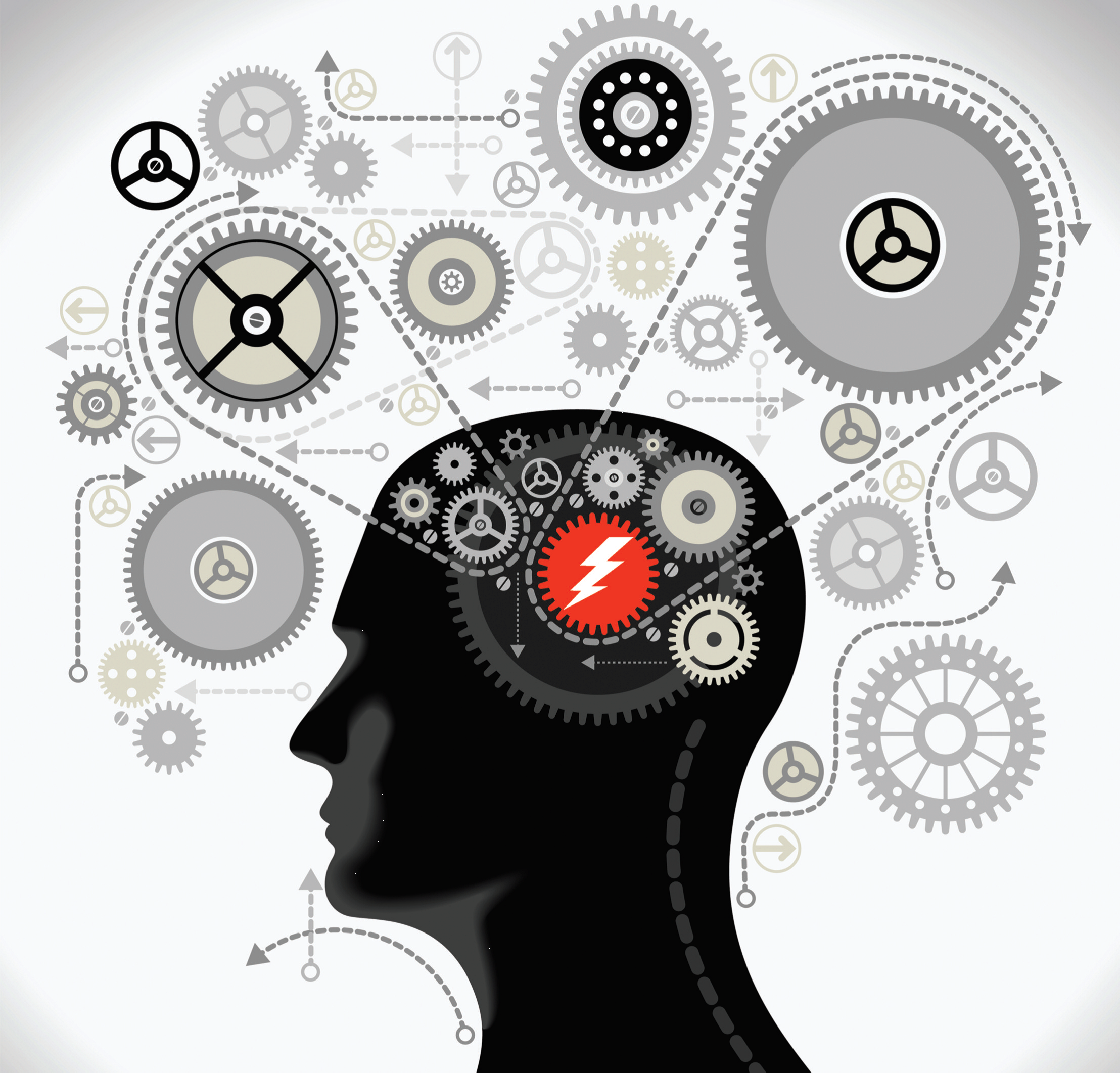
It is important for the epilepsy team to be well informed about the semiology of the seizures. Semiology means what the seizure looks like – how it starts, how it progresses, whether the patient is alert or not.
The word “seizure” strikes fear into the heart of parents. Nothing is more terrifying than watching your loved one suffer a convulsion, and even worse if it is your infant, child, or adolescent.
Seizures are more common in infancy and childhood than they are in adulthood, except in old age. The developing brain is particularly susceptible to seizures, which occur when the normal balance between excitation and inhibition is disrupted.
Seizures come in many shapes and sizes and, when occurring in childhood, are often self-limiting and fairly easily managed. Others may begin in childhood or adolescent and persist into adulthood but, again, are responsive to treatment.
However, some seizures are not so easily managed and become refractory or intractable. Some syndromes that occur early in childhood can persist and evolve into a lifelong disease.
Seizures that began in the neonatal period or in infancy may be quickly controlled, and the child taken off of medication, (in a few months to one to two years)—but other seizures that begin early on can be devastating and result in lifelong epilepsy and developmental delays.
Epilepsy is considered “refractory” (intractable) when it is not controlled with the first two medications, when used correctly with adequate blood levels and dosing. Most people (about 47 percent) become seizure free on the first medication that they are treated with. The chance of being well controlled on the second medication drops to about 13percent and with each subsequent medication, it drops even more.
The following article discuses some of the more well-known syndromes, but is by no means exhaustive.
WHAT IS EPILEPSY?
In general, epilepsy is diagnosed when a person has two or more unprovoked seizures— that is, seizures that are not caused by fevers, injuries, sleep deprivation, illness or drug or alcohol use. However, epilepsy may be diagnosed after only one seizure when the patient has an abnormal EEG.
The International League against Epilepsy (ILAE) has proposed a new definition for epilepsy which states that epilepsy be considered a disease of the brain defined by any of the following conditions:
1.At least two unprovoked seizures occurring more than 24 hours apart
2.One unprovoked seizure and a possibility of further seizures similar to the general recurrence risk after two unprovoked seizures (approximately 75 percent or more)
3.At least two seizures in a setting of reflex epilepsies (see below)
Every year about 450,000 children in the United States are newly diagnosed with epilepsy. Worldwide, there are about 10.5 million children who are diagnosed with epilepsy. The annual incidence of new cases of epilepsy (in adults and children) is 61 to 124 per 100,000 persons in developing countries and is somewhat lower in developed countries at 41 to 50 per 100,000 persons. (The difference is probably due to a higher incidence of injuries causing epilepsy in developing countries.) The annual rate of new cases of epilepsy is higher in children, with 100 to 200 new cases every year per 100,000 people children as opposed to 24 to 53 new cases each year for every 100,000 adults.
Some will become seizure free with medications, but 10 to 20 percent (400,000 to 800,000) will become medically intractable, meaning that their seizures cannot be controlled with medication. Of those with medically intractable seizures, one-third are surgical candidates (>5000 new per year). In children, about 10 to 20 percent will become medically intractable.
SYNDROMES THAT MASQUERADE AS EPILEPSY
It is important to differentiate between seizures and other conditions that mimic seizures. Some of the most common are syncope, breath-holding spells, benign paroxysmal vertigo, pseudo-seizures and episodic dys-control or rage attacks.
1.Breath-holding spells are an involuntary reflex that begins in toddlerhood and usually resolves by the time the child starts school. They are often triggered by an emotional response like anger, fright or frustration. The child cries, holds his or her breath and faints, sometimes while turning blue. The child can either be limp or stiff, or even shake.
2.Syncope or fainting is usually seen in older children or adolescents. The child may experience a warning sign such as blurred vision, lightheadedness, nausea or sweating. There may be some jerking at the end of the event and there is usually a quick recovery.
3.Pseudo-seizures are psychogenic or non-epileptic seizures. They are difficult to distinguish from epileptic seizures and are usually diagnosed after a video EEG captures the event. They occur with cases of psychiatric illness or emotional disturbances. They have also been referred to as “stress seizures”. Working with a psychiatrist is mandatory and has helped many patients. It is also important to note that 17 to 37 percent of patients with Pseudo-seizures have real Epilepsy too.
4.Episodic dyscontrol (rage attacks) are episodes of aggression out of proportion to the precipitating event. The child may not remember the explosive behavior that he or she exhibited (including hitting, kicking, biting.) They occur more often in children with autism, ADD or traumatic brain injury.
TYPES OF SEIZURES
The International League Against Epilepsy classifies epilepsies according to:
o Whether or not consciousness is altered
o The location of the disease in the brain
o The cause of the epilepsy
o And the main symptoms (what the seizures look like and how often they occur.)
Seizures are classified by their symptoms:
Dialeptic seizures include
• staring spells
• zoning out
• confused
Motor seizures include:
• Myoclonic seizures- quick jerking
• Tonic seizures- stiffening
• Clonic seizures- repetitive rhythmic jerking involving both sides of the body
• Tonic-clonic seizures- alternating jerking and stiffening
• Versive seizures- sustained, forceful turning
• Automotor seizures- automatisms of the hands, mouth or tongue, consciousness is usually compromised
• Spasms- sustained muscle contractions with flexion of the trunk and elevation of the arms
• Hypermotor seizures- large, violent movements Other seizure types:
• astatic or atonic seizures- loss of tone, with falls or head drops
• aphasic seizures- inability to speak
• hypomotor seizures- inability to move
• Sensory seizures- unusual feelings or sensations
• Autonomic seizures- piloerection, epigastic rising , bizarre heart racing
The older seizure classification system was based on whether they were partial (focal), denoting that they arise from one area of the brain, or generalized, arising from the whole brain.
Focal seizures included simple (no change in consciousness) and complex partial (patient was not alert.) Generalized seizures included myoclonic, absence, generalized tonic-clonic, and atonic, as well as spasms.
SEIZURES OCCURRING IN INFANCY
Seizures in infancy occur in 1.5 to 5.5/1000 newborns and most are focal; notable exceptions are myoclonic events and spasms,. Seizures in newborns can be quite subtle, including random and roving eye movements, or mouth movements like sucking, chewing and tongue protrusions or rowing, swimming or bicycling movements of the legs. Seizures in infancy are often caused by some provoking event such as infection, birth trauma, lack of oxygen, intracranial bleed or some metabolic derangement. Examples of metabolic derangement are chemical imbalances, such as low levels of magnesium, calcium, glucose or sodium in the blood. But there is a small collection of genetic disorders that present in infancy where the children quickly outgrow their seizures. They still need treatment and the diagnosis is made after a complete evaluation and by diagnosis of exclusion.
Seizures associated with vitamin B6 (pyridoxine) deficiency occur in the first 24 to 48 hours of life, but can occur as late as two to three years of age. They are often preceded by vomiting or illness and can present as spasms or groaning. They usually respond promptly to administration of intravenous vitamin B6.
Benign neonatal convulsions are focal clonic seizures or focal tonic seizures in a normal infant and no family history. Seizures occur at days four to six of life and are usually brief. They typically respond well to medication.
Benign familial neonatal convulsions (BFNC) are associated with a family history of neonatal convulsions and occur in an autosomal dominant pattern: potassium channel genes are associated with this disorder – KCNQ2 and KCNQ3. Seizures usually occur for two to three months, and then stop. They, too, typically respond well to treatment.
Benign myoclonic epilepsy is rare and more common in boys, about 40 percent of the time associated with a family history of febrile seizures or epilepsy. Seizures begin between five months and five years of age with head drops, eye deviation and arm jerks and the outcome is favorable.
Otohara syndrome (EIEE) is a severe epileptic encephalopathy occurring in early infancy. The EEG shows a distinct burst-suppression pattern and the infant suffers frequent seizures of a variety of types, including tonic, myoclonic and partial seizures. The syndrome has not been found to respond to any treatment, although some treatment options can slow certain types of seizures. It appears to have a few different genetic disorders causing it, but in most cases no clear genetic mutation has been found. No great treatment has been found yet, but one needs to try all the typical treatments that are currently available. It is associated with severe developmental disability and these patients require extensive care and the families need extensive support.
Early myoclonic epilepsy (EME) is characterized by myoclonus, usually in combination with other seizure types. The EEG shows a burst-suppression pattern. This infant encephalopathy has a much more dire outcome, and these seizures can be associated with an inborn error of metabolism, cerebral dysgenesis and hypoxic-ischemic insults.
Infantile spasms occur almost always in the first year of life and often do not respond to the usual antiepileptic medications. The seizures or spasms are flexion of the neck, trunk and extremities, but can also be more subtle, such as a shoulder shrug or eye roll. They can be associated with a cry and are very frequently mistaken for colic. Infantile spasms are also known as West syndrome, named for the triad of spasms, regression and EEG abnormalities. The EEG shows a chaotic background, with abnormal sleep and a hyppsarythmic pattern. EEG can also show a burst-suppression pattern. Infantile spasms are best treated with ACTH (Acthar Gel) or vigabatrin (SABRIL), and can evolve into Lennox-Gastaut syndrome, which continues into adulthood. Infantile spasms occur more frequently when there are malformations of the cerebral cortex or Tuberous Sclerosis or Down syndrome.
SEIZURES IN CHILDREN AND ADOLESCENTS
Febrile seizures are seizures that occur with fever or illness. They usually stop by the age of five and do not require treatment with preventative medication. They often run in families. There is a syndrome called GEFS (generalized epilepsy with febrile seizures plus) that occurs when children have febrile seizures beyond the age of five or six and then begin to have unprovoked seizures, including generalized tonic-clonic, absence, myoclonic and tonic. These disorders frequently (40 percent of the time) have a genetic mutation which can be found on special genetic testing.
Benign Rolandic epilepsy is also known as benign epilepsy with centro-rolandic or centro-temporal spikes . It is the most common idiopathic epilepsy, meaning that there is no associated brain abnormality or other obvious cause. Very often, children may have spikes on the EEG without actually having clinical seizures. Onset of the seizures usually occurs between the ages of three and 10 years old. There are usually no neurological or cognitive deficits. Most seizures are simple partial, meaning that there is no change in the level of consciousness, and the EEG tracings are characteristic. The BRE (benign rolandic epilepsy) seizures typically happen out of sleep , early in morning or at onset of sleep and typically involve the face and/or one side of the body. Sleep deprivation is the typical trigger. There are some genetic mutations associated with these disorders as well, and special testing is now available. BRE children also have a higher risk of learning issues and attention issues, so discuss the school aspects with your neurologists, not just the seizure issues.
Childhood absence epilepsy. Absence seizures used to be known as “petit mal” seizures. These occur as short episodes of loss of awareness that are associated with a distinct pattern on the EEG, a generalized spike and wave. These may be associated with a genetic abnormality affecting GABA A receptors and calcium channels. Affected genes are 5q34 and 19p. These are good examples of “Age Triggered Gene Defects”, in that the disorder turns on at a certain age and typically turns off after a few years (typically two to five years). Onset usually occurs between nine and 13 years of age.
Absence seizures can be typical or atypical. In typical absence epilepsy, most affected children have normal neurological examinations, although there is a high rate of learning disabilities and ADD. These seizures may rarely occur with a warning or aura and are brief. The person experiencing the seizures may only be aware of them when they notice that they may have lost time. These seizures usually respond to the first or second medication prescribed. The seizures may be associated with automatisms like lip smacking or fumbling. Atypical absence seizures may be longer than typical absence, and the automatisms may occur more frequently. There are more serious cognitive deficits associated with atypical absence.
Juvenile absence epilepsy has a peak onset between the ages of 9 and 13. The most common type of seizure is absence but generalized tonic-clonic seizures also occur in 80percent and myoclonic seizures may occur. These can be more difficult to outgrow and typically only 50 percent of children outgrow this disorder. However, it is usually reasonably well-controlled with medication.
Epilepsy with myoclonic absences occurs from five months to 12 years, with a peak incidence at seven years of age. Development is normal until seizures begin and the child has myoclonic absence seizures lasting from 10 to 60 seconds, occurring multiple times each day with myoclonus of the shoulders, and limbs.
Juvenile myoclonic epilepsy usually presents in adolescence or young adulthood, with peak onset between the ages of 14 and 16. Seizures often present as brief jerks (myoclonus) occurring upon awakening. Seizures are triggered by photosensitivity and sleep deprivation. Other generalized seizure types may also occur, including absence seizures and generalized tonic clonic seizures. Morning jerks often occur and generalized tonic-clonic seizures are often preceded by a series of jerks. JME frequently persists through adulthood, and typically is well controlled with medication. Most breakthrough seizures are due to teenagers refusing to be compliant with medication. Extra counseling is required here and nagging from parents.
Dravet syndrome or severe myoclonic epilepsy is a genetic syndrome that occurs when there is a mutation on the sodium channel gene SCN1A (the same gene defect discussed above in GEFS+). Seizures begin between the ages of two and 12 months and initially are generalized tonic-clonic or one sided tonic or tonic-clonic, are prolonged, and can be caused initially by fever and illness. Later in life, myoclonic seizures are the most common type, but other types, most commonly atypical absence, also occur. The child, who may have developed normally, begins to exhibit slowed development and the gait is awkward and often unsteady. The EEG may show fast spike and wave or polyspike and wave or can be multifocal. The background of the EEG becomes slower over the years. Anti-epileptic medications that work on sodium channels must be avoided since these can worsen the seizures. These include Carbamazepine (Tegretol) , oxcarbamezipine (Trileptal), lamotrigine (Lamictal), and phenytoin (Dilantin.)
ESES (epileptic status encephalopathy in sleep) or CSWS (continuous slow spike and wave in sleep) or Landau-Kleffner syndrome is a spectrum of syndromes that occurs in previously normally developing children, who suddenly experience a language regression. The language disturbances develop over a short period of time and are associated with behavioral disturbance in the majority of the children. The EEG shows bitemporal or posteriorly dominant spikes which occur very frequently during slow wave sleep. There are some cases where the child never has clinical seizures but because of the loss of language and the very abnormal EEG, this syndrome is still characterized as epileptic. When there are seizures, they are usually fairly easy to control, although the EEGs are much more difficult to control. Most cases remit before adulthood, but in 40 to 60 percent of cases, the child is left with a lasting language disorder involving difficulties with decoding sounds.
Myoclonic-astatic epilepsy (Doose syndrome) is characterized by myoclonic (jerks) and astatic (falls) seizures. Other types of seizures, including absence, may also occur. It occurs rarely, the structure of the brain is normal, and there is often a genetic component. Boys are affected more often than girls. Seizures usually begin between the ages of seven months and eight years, and most children develop normally until the seizures begin. The seizures occur many times each day and the EEG shows bursts of spikewave or polyspike and wave complexes. The course is unpredictable and may become intractable. These children frequently respond well to steroids or the Ketogenic diet. This diagnosis is frequently missed and confused with Lennox-Gastaut Syndrome. However, these children have a better outcome (elimination of seizures and improvement in development) if treated properly.
Frontal lobe seizures often occur out of sleep and may be mistaken for parasomnias. Seizures are often hypermotor, with wild thrashing about. The person may experience a change in the level of consciousness, and the seizures are usually brief with no postictal period.
Lennox-Gastaut syndrome is an epileptic encephalopathy with onset in childhood, featuring several types of seizures, including tonic, atonic, absence and sometimes myoclonic. These are associated with a distinct EEG pattern of slowing and spike and wave complexes. Patients affected are developmentally delayed or mentally retarded and may have cerebral palsy. Lennox-Gastaut syndrome is often the result of infantile spasms or other epileptic encephalopathies that begin in infancy. These seizures are multiple and difficult to treat, and require much communication between the family and medical team.
Occipital epilepsies can begin in early or late childhood. Panayiotopoulos syndrome is characterized by seizures that begin in early childhood and occur infrequently with vomiting and eye and head deviation, some progressing to generalized seizures. They usually remit after one to two years. The later onset occipital epilepsy is called Gastaut type and begins between the ages of eight and 11, remitting two to eight years, after onset in about 50 percent of patients. Seizures are very frequent, brief, with no loss of consciousness. They consist of simple visual auras with eye deviation or complex motor movements. They are frequently confused with migraines.
Stimulus sensitive epilepsies (Reflex Epilepsies) are invoked by specific stimuli and are also called reflex epilepsies. They include the following:
a. Photosensitive epilepsy
b. Seizures induced by complex activities (reading, music, particular voices)
c. Startles and movement induced epilepsy
d.Seizures produced by nonvisual stimuli
They occur rarely and their causes are poorly understood, and they respond moderately well to treatment.
Rasmussen’s Syndrome is a difficult to control focal epilepsy associated with inflammation, progressive cognitive decline and atrophy of one hemisphere of the brain. It usually presents between the ages of two and 14. It is treated with antiepileptic drugs, immunotherapies and hemispherotomy (surgical intervention.) They are typically associated with some illness, but sometimes the trigger is never found. One needs to be aggressive with treatment in these cases to help protect the good areas of the brain not yet affected by the epilepsy.
Some neurocutaneous (lesions on skin and brain) genetic syndromes are associated with seizures. These include the following syndromes:
Tuberous sclerosis – associated with skin lesions, tubers in the brain, abnormalities in the heart and kidneys and eyes, cognitive delays and autism. Seizures most often begin in infancy, are usually focal and often begin with infantile spasms. The genetic defects are either TSC1 or TSC2 which can be confirmed by blood tests.
Sturge Weber – associated with vascular malformations in the brain and port-wine stain facial birthmarks. This just recently is also able to be confirmed by genetic testing. Associated with very difficult to control seizures but all the children can have various degrees of severity of the underlying disorder.
Neurofibromatosis – associated with hypo and hyperpigmented birthmarks, fibromas, and learning disabilities. The Neurofibromin gene defect has been located on Chromosome 18. Epilepsy is seen in only 10 to20 percent of children of with NF1
Other genetic epilepsies include:
Hemimegalencephaly – increased size of one hemisphere of brain, responds to surgical intervention.
Rett syndrome – severe developmental delay, acquired microcephaly, occurs more often in girls, who develop normally until the age of one to two years old. Genetic defect is found on MECP2 gene and CDKL5 gene.
Ring chromosome 20 – seizures begin between three and five, the child may have mildly dysmorphic features, there may be mental retardation and cognitive decline.
Mitochondrial disorders – include MELAS and MERRF which are transmitted by the mother and intractable.
Cysticercosis – is the most common cause of epilepsy in Latin America and is caused by the ingestion of the eggs of Taenia solium, a tapeworm found in contaminated pork.
Hypothalamic hamartomas – are brain abnormalities that cause gelastic or laughing seizures and respond to surgical intervention. If not removed, they can cause cognitive decline.
POSTTRAUMATIC EPILEPSY
Seizures can occur after head trauma. In about one percent of concussions, there is an immediate seizure. With severe head injuries, (including bleeding in the brain and depressed skull fractures,) seizures may occur in the first week. This results in about a four times increase in the later risk of developing epilepsy. TBI (traumatic brain injury) patients can develop late onset epilepsy (occurring more than a week after the injury) which can often lead to chronic epilepsy. It is even possible that the seizures might not occur until up to four years after the injury.
IN CONCLUSION
It is important for the epilepsy team to be well informed about the semiology (the description) of the seizures. Semiology means what the seizure looks like – how does it start, how does it progress, whether the patient is alert or not. We have listed here some websites that give guidelines for seizure diaries, as well as some with information regarding seizures and epilepsy.•
ABOUT THE AUTHORS:
Steven M. Wolf, M.D., is a Pediatric Neurologist at Ferncliff Manor, and Director of the Developmental Disability Center at Roosevelt Hospital and the Director of Pediatric Epilepsy at Beth Israel Medical Center and EEG Lab at St. Luke’s-Roosevelt Hospital Center. His practice is in collaboration with Patricia McGoldrick, NP.
Patricia Engel-McGoldrick, RN, is a Nurse Practitioner and a Medical Physician Assistant, she is also Associate Director of the Developmental Disability Center at St. Luke’s Roosevelt Hospital and Co-director of the Tuberous Sclerosis Clinic at Beth Israel Medical Center.
References
i Kwan and Brodie. Early Identification of Refractory Epilepsy 2/3/2000 342:314-31 NEJM
ii Guerrini R. Epilepsy in children. Lancet 2006:367(9509):499-524
iii Wylie E. Catastrophic epilepsy in infants and children: Identification of surgical candidates. Epileptic Disorders 1999;1(4):261-264
iv Duchowny M. Epilepsy surgery in children. Curr Opin Neurol 1995;8(2):112-116
v Wylie E. et al. Seizure outcome after epilepsy surgery in children and adolescents. Ann Neurol
1998;44(5):740-748
vi Luders H. Lesser RP, Dinner DS et al 1987 Benign focal epilepsy of childhood In: Luders H, Lesser RD, eds. Epilepsy: Electroclinical Syndromes. Berlin: Springer, 303-346.
vii Lisa Strug & Deb Pal 2010
viii Jennett B et al Epilepsy after head injury Br Med Journal 1978 July 22;2(6132):229